PHOTO GALLERY
Spectral Images of Normal and Cancerous Breast Tissue click here
CD Ratiometer click here
Artist's Rendering of Sarnoff-MTC Tissue Fluorescene Imager click here
Spectral Images of Normal and Cancerous Breast Tissue
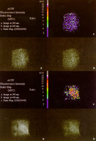
Top:Spectral images of normal breast tissue excited at 300nm, having a ratio
of 2.5.(a)Fluorescence image for wavelength at 340nm, (b) Flourescence image
for wavelength at 440 nm, (c) Ratio map with a discrete pallet.
Bottom: Spectral images of cancerous breast tissue excited at 300 nm, having
a ratio of 10.5. (a) Fluorescence image for wavelength at 340 nm, (b)
Fluorescence image for wavelength at 440nm, (c) Ratio map with a discrete
pallet.
Top of page
Prototype CD Ratiometer
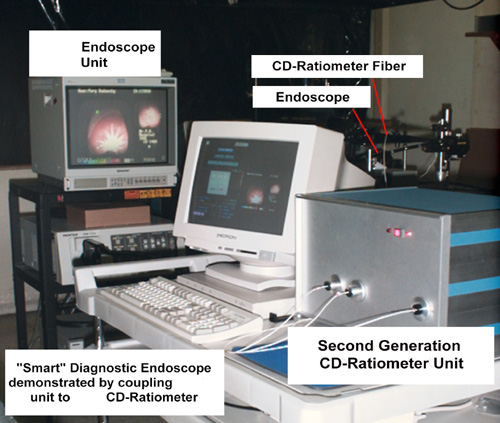 The CD Ratiometer designed to optically assess the scanned tissue only at pre-established
optical wavelengths and to instantaneously report out a "yes" "no" or "maybe"
viewed on a crt screen. Click here to read more. Top of page
Artist's Rendering of Sarnoff-MTC Tissue Fluorescene Imager

As seen the scope can be used in conjunction with
other viewing devices such as an endoscope and microscope.
Top of page
|