|
The New York State Center for Advanced Technology ULTRAFAST PHOTONIC MATERIALS AND APPLICATIONS The City University of New York and The Center for Laser Imaging and Cancer Diagnostics A US Department of Energy-Supported Center of Excellence for Lasers in Medicine
Originally Produced for Breast Cancer and Women�s Health Awareness Day, sponsored by the New York State Legislative Commission on Science and Technology, Albany, NY, March 11, 1998 � 1998 V. Tomaselli, R. Alfano, S. Gayen, A. Katz, Q.Z. Wang, P.-P. Ho, K. Sutkus 1. Introduction Optical biopsy and optical imaging are two emerging complementary photonic technologies devoted to the use light to diagnose diseases and peer inside human body in search of lesions. This brochure presents a brief introduction to the photonic technologies under development at the City College of the City University of New York, that are expected to be safe, noninvasive, and more affordable with genuine diagnostic ability.
Light interacts with biological tissues through a variety of processes that include reflection, refraction, absorption, emission, as well as elastic and inelastic scattering. Optical biopsy techniques use the 'color of light', that is, the spectroscopic differences between normal and cancerous tissues to diagnose the disease. The difference in light transmission through normal and infected tissues provides the physical basis for optical imaging. A combination of these two major approaches is expected to provide genuine diagnostic imaging modalities. The interest in developing light-based medical imaging and diagnostic techniques derives from several favorable characteristics. First, light is a less-invasive and non-ionizing radiation so that routine screening with light poses no health hazard. Second, in contrast with available techniques, optical biopsy do not require removal of tissues, and the diagnosis is in situ and rapid. Third, optical imaging techniques have diagnostic potential, use of light of different wavelengths enables exploitation of spectroscopic signatures. Optical techniques are expected to be simpler and more affordable than the existing methods. Optical biopsy and optical imaging techniques currently under development at CCNY cover a wide range of diseases and body organs. The focus of optical biopsy techniques is to diagnose cancers with high accuracy and specificity. Major thrust of optical imaging research is to develop screening modalities for breast and prostate cancers. The techniques are in different stages of development. Prototype instruments developed for fluorescence-based cancer diagnosis are undergoing FDA-approved testing. Some of the imaging techniques are being tested on excised tissue specimens. Patents have been secured to protect intellectual property rights. Researchers at CCNY are continually striving to make novel photonic technologies available to medical practitioners. The remainder of the brochure is organized as follows. Section 2 introduces the organizational structure of photonic biomedical research efforts at CCNY. Section 3 highlights the diagnostic instruments that have been developed. Sections 4 provides a general background of optical methods in medical biopsy and imaging. The basic principles and considerations involved in imaging objects inside turbid media, such as tumors in breast, are discussed in Section 5. Section 6 presents the basics of fluorescence spectroscopy and its application in cancer diagnosis. The brochure ends with several appendices that define commonly used terminology, list selected publications and patents by CCNY researchers, and refers to literature for further reading.
2. The City College Institute for Ultrafast Spectroscopy and Lasers Founded in 1982 to oversee the various ongoing research studies, the Institute (IUSL) is the parent organization for several organizations that house and support research on lasers, optical materials, systems and applications such as tunable solid-state lasers, optical imaging in turbid media, and medical diagnostics. Achievements in these projects led to the growth and expansion of the effort, scope, and funding of IUSL research. Today, the IUSL serves as an umbrella organization for three major programs at the City University of New York (CUNY). 2.1. The New York State Centers for Advanced Technology Since 1983, major research universities throughout New York State have worked with industry partners on problems of mutual interest, and benefit to the state�s economy, through the Centers for Advanced Technology (CAT) Program. The 13 CATs, located at major research institutions in New York State, each support a different area of specialization, but share a common goal of technology transfer and commercialization of their results. The CAT program provides industry access to the innovative technology and facilities available at the participating research institutions. The CAT in Ultrafast Photonic Materials and Applications at CUNY, which began operations in 1994, supports several areas of research and technology development:
2.2. Institutional Research Award Funded from the NASA Office of Minority University Research and Education Division (MURED) to study Tunable Solid-State Lasers and Optical Imaging, the IRA program promotes the dual roles of cutting-edge research in areas that support NASA programs and providing meaningful research education and training for minority students, primarily Hispanics. Awarded in 1994, this 5-year, $3.8 million program encompasses faculty and students from 4 of CUNY�s senior colleges.
2.3. Center for Laser Imaging and Cancer Diagnostics Following a highly competitive application process, the US Department of Energy, Office of Energy Research awarded a Center on Laser Imaging and Cancer Diagnostics to City College. The proposed approach builds on existing technologies and partnerships with major medical research centers in New York to detect breast and prostate cancer. Partners in this new Center include Memorial Sloan-Kettering Cancer Center, New York Hospital-Cornell Medical School, Hackensack University Medical Center, and Englewood Hospital, as well as Lawrence Livermore Laboratory, a National Laboratory. An industrial advisory board, consisting of representatives from major medical instrument manufacturers, is an integral part of the Center�s structure.
3. Research Program Medical diagnostics research at City College is mainly involved with the detection of cancer using several optical-based techniques. These techniques have, to a large extent, been pioneered at CUNY over the last 10 years. At the City College, Director Robert Alfano and co-workers have invented optical techniques for dental caries detection, cataract monitoring, cancer detection, and optical counterparts to mammography. Since 1984, medical diagnostic research has been focused on the characterization of tissue to distinguish normal, benign, and cancerous samples. This work has led to the development of two instruments - CD Scan and CD Ratiometer. (CD means cancer detection). This pamphlet describes some of the basic elements underlying the operation of the CD instruments.
For further information, contact: Dr. Robert R. Alfano Distinguished Professor of Science and Engineering Director, Institute for Ultrafast Spectroscopy and Lasers Director, Center for Advanced Technology City College of CUNY Convent Avenue at 138th Street New York, NY, 10031 voice 212.650.5533; fax 212.650.5530. email: [email protected]
Dr. Vincent P. Tomaselli Deputy Director, Business Development and Operations Center for Advanced Technology Research Foundation CUNY 30 West Broadway, 11th Floor New York, NY 10007 voice: 212.417.8320; fax: 212.417.6448 email: [email protected]
4. Background Breast cancer is the leading cause of death in American women from 40-49. It is generally accepted that early detection is an effective means to minimize adverse effects. In women over 50, there is a 30% drop in death rates from breast cancer for those having regular mammograms However, a recent (January 1997) panel, convened by the National Institutes of Health, concluded that for women in their 40�s, annual mammograms, while possibly preventing death in a small percentage of the population, might cause harm from exposure to radiation (x-rays), encouragement of unnecessary surgery, or false complacency. In N1996, researchers at the University of Alabama at Birmingham published a study in the journal CANCER (1996: 78:2045-8) reporting that for the first time in recorded US history, cancer death rates, for the period 1990-1995, declined. The total reduction in all cancers was 3.1%. Not surprisingly, declines in breast cancer death rates were attributed to early detection and improved treatment. Thus, early, sensitive tumor screening is effective, especially for women over 50. But for younger women, off-setting benefits, including exposure to ionizing radiation, make the choice of the standard x-ray mammography screening protocol less clear.
4.1. Alternate Diagnostic Technologies Research efforts to find cancer detection methods, based on alternate technologies, have been developed. In addition to X-RAY MAMMOGRAPHY, which is not well-suited for younger women, and which may not detect early-stage tumors, there are several other techniques, each of which has advantages and disadvantages.
4.2. Optical Methods Optical methods in mammography and biopsy are emerging as effective, alternate cancer detection techniques. Recent research progress has shown that optical methods can be safe, sensitive, reliable, and reasonably cost-effective. A number of optical techniques are being developed by scientists worldwide, and progress is encouraging.* *On April 24-25, 1997, the CUNY CAT and the New York Academy of Sciences co-sponsored a 2-day symposium on Advances in Optical Biopsy and Optical Mammography. The proceedings (Annals, Vol. 838) are available from NYAS (2 East 63rd Street, New York, NY 10021; 212.838.0230, Ext. 324). Medical optical diagnostics research at the City College/IUSL has focused on two areas Studies on OPTICAL DETECTION have resulted in instruments that can be used to screen for cancer. Preliminary laboratory tests have been completed on one of two devices, and FDA-approved testing is in progress. Optical imaging is still in the basic research stage. Functioning, clinic-ready instruments that produce a clear image of a tumor are a few years away. In this pamphlet, some ideas to help the reader understand optical methods are discussed.
5. Basics of Optical Imaging in Turbid Media - Toward Optical Mammography A shadowgram is a projection of an illuminated object onto a screen, much as a slide or transparency taken with a camera is projected onto a screen. The problem in mammography is to detect a tumor within the breast by viewing its captured image. As with normal photography, an image results from tissue density differences. The characteristics of the projected image allows the diagnostician to evaluate the state of the object (tumor). For simple shadowgrams (normal photography), the medium surrounding the object is transparent, allowing an image to easily be formed. Detecting a tumor within breast tissue is somewhat more complicated. First, the surrounding medium (breast tissue) is not transparent, but is highly turbid. This turbidity results in the loss of image information as the illuminating light travels through the tissue. Second, the density differences between the normal tissue and tumor can be small resulting in low contrast, and consequently a faint image. A glass of milk is an example of a turbid medium. An object (such as a spoon or straw) cannot be seen if it is in the center of the glass of milk because the image-carrying information has been destroyed by the light scattering caused by the suspended milk particles. Without eliminating the turbid medium or compensating for the image-degradation it has caused, an imbedded object is not detectable (visible). 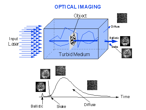
CAT researchers, studying the properties of turbid media, found that if the illuminating light is a short-lived (ultrafast) laser pulse, the resulting light transmitted through the medium has three components called ballistic, snake, and diffuse. The ballistic component is the light that is least scattered out of the original beam direction and, consequently, reaches the projection screen first. It contains useful image information and forms a sharp image. The problem is that ballistic light is very weak compared to the other
components, so that the image it forms is very faint. The largest component of the three is diffuse light and, as might be expected, carries no useful image information with it. In effect all image-forming information is lost as the light undergoes multiple encounters (scattering) with the turbid medium. Snake light is the component that is scattered off-axis more than the ballistic component, arrives at the screen or detector later than ballistic light, but sooner than the predominant diffuse light. Snake light contains some useful image information, and separates the other two regions. If a useful image is to be extracted from the total light passing through the object and surrounding medium, it is necessary to capture the ballistic light and filter out the non-imaging components. Time-gating is a technique in which a light valve or gate is opened for an extremely short period of time to let pass the desirable light, and filter out undesirable light. A time gate serves the same role as a shutter in a camera. Using time-gating detection, CUNY CAT researchers are able to filter out the ballistic and snake light and prevent the image-destroying diffusive light from reaching the screen. Figure 1 is a schematic diagram of an object (2-dimensional line drawing of a cat) that is immersed in a turbid medium. The image formed of the cat by the ballistic component is sharp. That formed by the diffuse component is not decipherable. The snake light image is blurred, but recognizable. 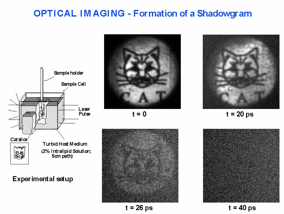
For this gating method, time intervals are measured in units of picoseconds (1 ps = 10-12
seconds, or 1/1,000,000,000,000 seconds). In Figure 2, more details of the experimental set-up are shown. The four images of the cat are shown for differing gate times, that is times when light is allowed to pass through (open shutter). The t=0 ps image, formed by ballistic light, produces a sharp image. If the gate is opened for t=20 ps, the image is formed by both ballistic and snake light, and is showing some degradation introduced by the snake light. At t=26 ps, the time gate is open longer allowing more snake and some diffuse light to pass. The result is a barely discernible image. At t=40 ps, the diffusive component has become dominant and no useful information is captured by the imaging system - the cat has disappeared. In summary, with ultrafast lasers and time-gating detection methods, it is possible to pick out very weak images of objects immersed within diffuse, cloudy media. This same experimental set-up is under development to enable imaging of tumors in breast tissue. The concept was invented and developed at City College in 1991.
6. Basics of Fluorescence Spectroscopy - Toward Optical Biopsy A material that is illuminated with light of a certain wavelength (color) and then re-emits light at the same or different wavelength is said to fluoresce. The collection and analysis of all re-emitted light from the excited sample is called fluorescence spectroscopy. Biological molecules such as collagen, elastin, NADH (a nucleotide involved in cell energetics), and tryptophan, substances found in human and animal tissue, fluoresce. The chemical composition and structure of thbiological substances determine the characteristics of the fluorescence spectrum. If normal and diseased biological tissue are spectroscopically different, and the difference(s) can be distinguished using fluorescence, a detection scheme is possible. 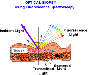
To create a fluorescence spectrum, the tissue sample is illuminated with light from a lamp (steady, broad-band source). This incident light can pass through the tissue (transmitted), be scattered internally, or re-emitted as fluorescence light (see Figure 3). To analyze the fluorescence light, collection optics (fibers, mirrors, filters, etc.) direct the light from the sample to an analyzer and then to a detector and output device which senses and displays a graph of light intensity vs. wavelength, that is, the spectrum. Cancer detection using optical fluorescence relies on the fact that normal and cancerous tissue have native fluorescence spectra that are different. In Figure 4, a fluorescence spectra from in vitro samples of cancerous and benign tissue are shown. The spectra, recorded over the range from 320 nm (ultraviolet) to 580 nm (yellow), show small, but clear differences between the spectral features of the two samples. These differences can be use to identify cancerous tissue. In practice, to quantify the process and make the diagnosis amenable to automated analysis, the ratios of intensities at selected wavelengths are used to provide a sample signature. In the case shown in Figure 4, the ratio of the intensities at 340 nm and 440 nm for benign and normal tissue generally fall below �9�, while that for cancerous tissue is generally over �9�. The particular value of the ratio (9) is not critical, and can, in fact, change at other wavelengths. However, the clear distinction between the two classes of tissue is important, and is the key to using fluorescence spectroscopy to detect cancer. 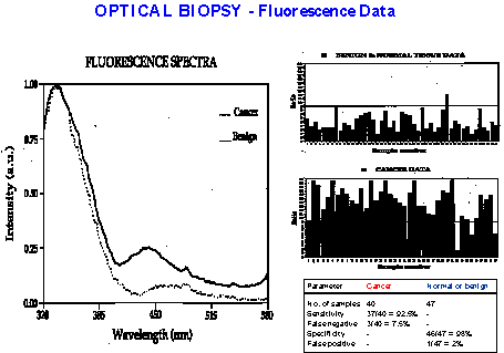
The table in Figure 4 lists useful parameters to evaluate the effectiveness of a diagnostic procedure. For the same sampling discussed above, the sensitivity (should be high) in detecting cancer in 40 samples is 92%, while the incidence of false negatives (should be small) is 7.5%. Normal sample parameters are similarly encouraging (low incidence of false positives - 2%- and high specificity - 98%). These parameters compare very favorably to normal pathology.
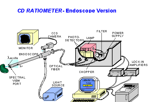
6.1. CD Ratiometer Using fluorescence spectroscopy, researchers at City College have developed two versions of an instrument called the CD RATIOMETER. Schematic diagrams of these devices are shown in Figures 5 and 6. In the endoscope version (Figure 5), an optical fiber carries exciting light (labeled l excite ) from a source (lamp) to a sample through an endoscope. The sample under examination re-emits light (l F) that travels back through the optical fiber. A series of filters is used to select the wavelengths that reach the photodetectors and that are used to compute the characterizing intensity ratio. The result is displayed on a computer monitor, and can be printed out for a permanent record.
Figure 5 The endoscope version of the CD RATIOMETER fiber can be used in combination with a co-linear viewing fiber that captures a video image (CCD camera) and presents it on a video monitor. This allows the diagnostician to visually identify a region of tissue for examination. Once the tissue area is visually selected, the CD RATIOMETER can be used to characterize it optically. The needle version concept shown in Figure 6 operates on the same principle as does the endoscope version. However, for this invasive method, a smaller optical fiber is needed. 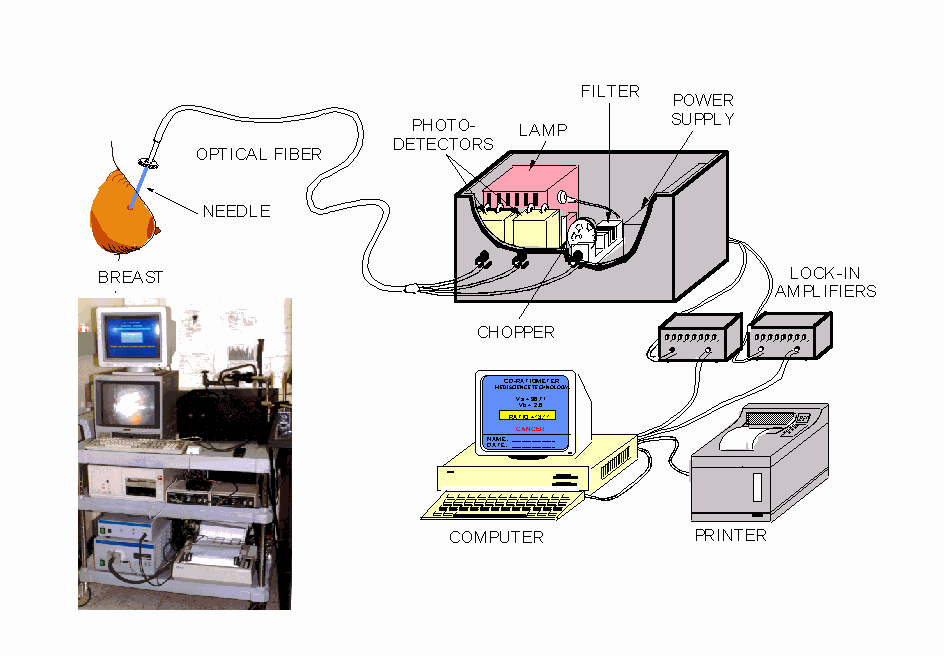
7. Research Results The medical diagnostics research using optical methods, performed by CAT Director Alfano and co-workers at City College of CUNY since 1984, is extensive. Many of the studies are pioneering and have resulted in significant contributions to the field. Appendix B is a representative list of the publications that have resulted from this work. These research and development studies have been supported by major organizations and agencies such as the New York State Science and Technology Foundation, National Institutes of Health, US Navy, US Air Force, National Aeronautics and Space Administration, and Ballistics Missile Defense Command. In addition, industry support, mainly from Mediscience Technology Corp., has been received.
Appendix C is a list of patents that have been received for devices and procedures applicable to medical diagnostics and related areas. This list includes patents covering the devices described above for instruments used in optical biopsy and optical mammography. The technology covered by these patents are available for commercialization. For medical use, FDA approval is required. � 1998 V. Tomaselli, R. Alfano, S. Gayen, A. Katz, Q.Z. Wang, P.-P. Ho, K. Sutkus Appendix A Glossary Photonics - Technology that uses the photon as a carrier of information; an analogue to electronics where the information carrier is the electron. Includes light generation, emission, transmission, deflection, amplification and detection by optical components and optical instruments, lasers and other light sources, fiber optics, electro-optical instrumentation, and related hardware systems.Mediphotonics - The application of photonic technology and science to medicine, covering uses ranging from diagnostics to therapy. Applications of mediphotonics include imaging, analysis, diagnosis and treatment of biomedical samples through optical technology. Optical Spectroscopy- The study of the intensity profile of a material as a function of wavelength. The data trace is called an optical spectrum, and it arises from absorption, emission or scattering of incident light. Optical spectra result from the interaction of a material with an electromagnetic field and are indicative of basic physical, biological and chemical characteristics of the sample material. Optical Biopsy - A medical diagnostic technique which makes use of optical spectroscopy to determine the state of a biological sample. Optical Mammography -The use of optical techniques to image the interior of the human breast and display the result in the form of a shadowgram for medical diagnostics. Optical Tomography - The combination of optical techniques, mathematical algorithms, and computer technology to produce three-dimensional images of the interior of objects hidden within scattering media. Ballistic Photons - Optical signals resulting from the coherent interference of light scattered in the forward direction that propagates straight through the medium. Ballistic photons travel along the minimum path or the shortest time to arrive at the detector in the forward direction. Snake Photons - Photons which travel along a zig-zag path, deviating slightly from the original input direction, and arriving at a detector later than ballistic photons. Snake photons are the early-arriving detected light traveling through a scattering medium in the near forward direction, which arrive within the first 30-ps of the event. They are quasi-diffusive photons - they do not obey the diffusion statistics. Diffusive Photons - Photons which follow a random walk path through the scattering medium, and arrive at a detector �late�, and with a broad temporal distribution. Diffusive photons constitute the largest component of scattered light and are responsible for the loss of image information in transillumination. Inverse Scattering Problem - The use of mathematical algorithms to reconstruct a map of the interior of an object by analyzing the diffusive light scattered by the medium. Ballistic Time - The time needed for a ballistic photon to travel a distance (L), in a medium. Mathematically, it is represented by the equation T = L/cn, where cn is the speed of light in the medium (c/n; n= index of refraction of the medium). Picosecond - A unit of time that is convenient for measuring fast processes. Abbreviated ps, where 1 ps = 10-12 seconds. Light travels 300 microns (a few hair widths) in one picosecond. Femtosecond - A unit of time that is convenient for measuring very fast processes. Abbreviated fs, where 1 fs = 10-15 seconds. Light travels .3 microns in one femtosecond. Gating - A technique for selecting out parts of a light signal profile. Can be time-gated or space-gated. Gating allows for the exclusion of parts of the signal that cause degradation of image quality. Appendix B Publications on Optical Biopsy and Optical Mammography Optical Mammography: Time-Resolved Degree Of Polarization For Human Breast Tissue, S. G. Demos, H. Savage, A. S. Heerdt, S. Schantz, R. R. Alfano, Optics Commun. 124, 439 (1996). Temporal Gating in Highly Scattering Media by the Degree of Optical Polarization, S. G. Demos, R. R. Alfano, Opt. Lett. 21, 161 (1996). Time-Resolved Optical Imaging of Jet Sprays and Droplets in Highly Scattering Medium, P. A. Galland, X. Liang, L. Wang, K. Breisacher, L. Liou, P. P. Ho, R. R. Alfano, ASME, HTD-Vol. 321, 585 (1996). Emerging Optical Biomedical Imaging Techniques, S. K. Gayen, R. R. Alfano, Optics & Photonics News, p. 17, March (1996). Imaging of Microscopic Objects Hidden behind a Highly Scattering Media, F. Liu, Y. X. Shan, R. R. Alfano, SPIE 2387, 204 (1995). Fourier Spatial Filter acts as a Temporal Gate for Light Propagating through a Turbid Medium, Q. Z. Wang, X. Liang, L. Wang, P. P. Ho, R. R. Alfano, Opt. Lett. 20, 1498 (1995). Two-dimensional Kerr-Fourier Imaging of Translucent Phantoms in Thick Turbid Media, X. Liang, L.. Wang, P. P. Ho, R. R. Alfano, Appl. Opt. 34, 3463 (1995). True Scattering Coefficients of Turbid Matter Measured by Early-time Gating, L. Wang, X. Liang, P. Galland, P. P. Ho, R. R. Alfano, Opt. Lett. 20, 913 (1995). Spectral and Temporal Measurements of Laser Action of Rhodamine 640 Dye in Strongly Scattering Media, W. L. Sha, C.-H. Liu, R. R. Alfano, Opt. Lett. 19, 1922 (1994). Microscope Imaging through Highly Scattering Media, G. E. Anderson, F. Liu, R. R. Alfano, Opt. Lett. 19, 981 (1994). Time-resolved Imaging of Translucent Droplets in Highly Scattering Turbid Media, R. R. Alfano, X. Liang, L. Wang, P. P. Ho, Science 264, 1913 (1994). Transmitted Photon Intensity through Biological Tissues within Various Time Windows, F. Liu, K. M. Yoo, R. R. Alfano, Opt. Lett. 19, 740 (1994). IR Fourier Space Gate and Absorption Imaging through Random Media, J. J. Dolne, K. M. Yoo, F. Liu, R. R. Alfano, Lasers in the Life Sciences 6(2), 131 (1994). Should the Photon Flux or the Photon Density be used to Describe the Temporal Profiles of Scattered Ultrashort Laser Pulses in Random Media?, F. Liu, K. M. Yoo, R. R. Alfano, Opt. Lett. 18, 432 (1993). Ultrafast Time-gated Imaging in Thick Tissues: A Step Toward Optical Mammography, B. B. Das, K. M. Yoo, R. R. Alfano, Opt. Lett. 18, 1092 (1993). Double-stage Picosecond Kerr Gate for Ballistic Time-gated Optical Imaging in Turbid Media, L. M. Wang, P. P. Ho, R. R. Alfano, Appl. Opt. 32, 535 (1993). Ultrafast Laser-pulse Transmission and Imaging through Biological Tissues, F. Liu, K. M. Yoo, R. R. Alfano, Appl. Opt. 32, 554 (1993). Spectral Optical-density Measurements of Small Particles and Breast Tissues, B. B. Das, K. M. Yoo, F. Liu, J. Cleary, R. Prudente, E. Celmer, R. R. Alfano, Appl. Opt. 32, 549 (1993). Imaging of a Translucent Object hidden in a Highly Scattering Medium from the Early Portion of the Diffuse Component of a Transmitted Ultrafast Laser Pulse, K. M. Yoo, B. B. Das, and R. R. Alfano, Opt. Lett. 17, 958 (1992). Optical Imaging vs X-rays for Breast Cancer Screening, R. R. Alfano, P. P. Ho, K. Yoo, Photonics Spectra, pp109-114, Oct. 1992 Ballistic 2-D Imaging Through Scattering Walls Using an Ultrafast Optical Kerr Gate, L. Wang, P. P. Ho, C. Liu, G. Zhang, R. R. Alfano, Science 253, 769 (1991). Speed Of The Coherent Component Of The Femtosecond Laser Pulses Propagating Through Random Scattering Media, F. Liu, K. M. Yoo, R. R. Alfano, Opt. Lett. 16, 351 (1991). Imaging through a Scattering Wall using Absorption, K. M. Yoo, F. Liu, R. R. Alfano, Opt. Lett. 16, 1068 (1991). When does the Diffusion Approximation Fail to Describe Photon Transport in Random Media?, K. M. Yoo, F. Liu, R. R. Alfano, Phys. Rev. Lett. 64, 2647 May (1990). Time Resolved Coherent and Incoherent Components of Forward Light Scattering in Random Media, K. M. Yoo, R. R. Alfano, Opt. Lett. 15, 320 (1990). Optical Biopsy Laser Induced Fluorescence Spectroscopy from Native Cancerous and Normal Tissue, R. R. Alfano, D. B. Tata, J. Cordero, P. Tomashefsky, R. W. Longo, M. A. Alfano, IEEE J. of QE 20 ,12 p. 1507(1984) Fluorescence Polarization Spectroscopy and Time-Resolved Fluorescence Kinetics of Native Cancerous and Normal Rat Kidney Tissues, D. B. Tatta, M. Foresti, J. Cordero, P. Tomashefsky, M. A. Alfano, R. R. Alfano, Biophysical J., Vol. 50 p463 (1986) Fluorescence Spectra from Normal Human Breast and Lung Tissues R. R. Alfano, G. C. Tang, A. Pradhan, W. LAm, D. S. J. Choy, E. Opher, IEEE J. of QE Vol.23, 10 p1806 (1987) Steady State and Time-Resolved Laser Fluorescence from Normal and Tumor Lung and Breast Tissues , R. R. Alfano, G. C. Tang, A. Pradhan, M. Bleich, D. S. J Choy, E. Opher ,J. of Tumor Marker Oncology Vol.3 p165 (1988) Spectroscopic Differences Between Human Cancer and Normal Lung and Breast Tissues , G. C. Tang, A. Pradhan, R. R. Alfano, Lasers in Surgery & Med. p290 1989 Optical Spectroscopic Diagnosis of Cancer and Normal Breast Tissues , R. R. Alfano, A. Pradhan, G. C. Tang, S. J. Wahl JOSA B 6 5 1015 5 1989 Pulsed and CW Laser Fluorescence Spectra from Cancerous, Normal and Chemically Treated Normal Human Breast and Lung Tissues , G. C. Tang, A. Pradhan, W. Sha, J. Chen, C. H. Liu, S. J. Wahl, R. R. Alfano, Applied Optics Vol. 28 12 p2337 1989 Effects of Self-Absorption by Hemoglobins on the Fluorescence Spectra from Normal and Cancerous Tissues, C. H. Liu, G. C. Tang, A. Pradhan, W. L. Sha, R. R. Alfano, Lasers in the Life Sciences, Vol. 3 No.3 p167 (1990) Human Breast Tissues Studied by IR-Fourier Transform Raman Spectroscopy , R. R. Alfano, C. H. Liu, W. L. Sha, H. R. Zhu, D. L. Adkins, J. Cleary, R. Prudente, E. Cellmer, Lasers in the Life Sciences Vol.4 No. 1 p23 (1991) UV Fluorescence Spectroscopic Technique in the Diagnosis of Breast, Ovarian, Uterus, and Cervix Cancer B. Das, W. Sha Glassman, R. R. Alfano, J. Cleary, R. Prudente, E. Celmer, S. Lubicz,,SPIE Vol.1427 p368 (1991) Light Sheds Light on Cancer-Distinguishing Malignant Tumors from Benign Tissues and Tumors, R. R. Alfano, B. B. Das, J. Cleary, R. Prudente, E. Celmer. B. of the NY Acad. of Sci. Vol.67 No.2 p143 (1991) Raman, fluorescence and time-resolved light scattering as optical diagnostic techniques to separate diseased and normal biomedical media , C. H. Liu, B. B. Das,et al, R. R. Alfano, J. Photochem. Photobiol. Vol.16 p187 (1992) Time Resolved UV Photoexcited Fluorescence Kinetics from Malignant and Non-Malignant Human Breast Tissues , A. Pradhan, B. B. Das, K. M. Yoo,J.Cleary,R.Prudente,E.Celmer,,R.R.Alfano, Lasers in the Life Sciences Vol.4 No.4 p225 (1992) Ultraviolet Excited Fluorescence Spectra from Non-malignant and Malignant Tissues of the Gynecological Tract W. Sha Glassman, C. H. Liu, G. C. Tang, S. Lubicz, R. R. Alfano, Lasers in the Life Sciences Vol. 5 No. 1 p49 (1992) Native Fluorescence Spectroscopic Detection of the Effects of Chemotherapeutic Retinoids on a Cancer Model of the Aero-digestive Tract, G.C.Tang, H.E.Savage,M.Silverberg,P.G.Sacks,V.Reid,S.P.Schantz,R.R.Alfano, Optics for Protection of Man and Environment p205 (1993) Native Fluorescence Spectroscopy of Thymus and Fat Tissues , G.Tang, M.Oz, V. Reid,R. Alfano, et al, SPIE Vol. 1887 p165 (1993) NIR Raman and Fluorescence Spectroscopies Diagnose Cancer , C.Liu, B.Das, W. Sha, G.Tang, H.Zhu, D. Akins, R.Alfano, SPIE Vol. 1887 No.189 (1993) Time Resolved and Steady State Fluorescence Spectroscopy from Normal and Malignant Cultured Human Breast CellLines , W. S. Glassman, M. Steinberg,,R. R. Alfano, Lasers in the Life Sciences Vol. 6 No. 2 p91 (1994) Excitation Spectroscopy of Malignant and Non-malignant Gynecological Tissues , W. Glassman, C. H. Liu, S. Lubicz,R. R. Alfano Lasers in the Life Sciences Vol. 6 No. 2 p99 (1994) Optical Spectroscopy Methods to Detect Colon Cancer , Y. Yang, G. C. Tang, M. Bessler, R. R. Alfano, SPIE Vol. 2135 p36 (1994) Fluorescence Diagnosis of Gynecological Cancerous and Normal Tissues , Z. Z. Huang, W. Sha Glassman, G. C. TangS. Lubicz, R. R. Alfano, SPIE Vol. 1235 p42 (1994) Tissue Autofluorescence as an Intermediate Endpoint in NMBA-induced Esophogeal Carcinogenesis, R. Glasgold, M. Glasgold, H. Savage,J. Pinto, R. R. Alfano, S. Schantz, Cancer Letters Vol.82 p33 (1994) Detecting Retinoic Acid-Induced Bio-chemical Alterations in Squamous Cell Carcinoma Using Intrinsic FluorescenceSpectroscopy , M. B. Silberberg, H. E. Savage,G. C. Tang, P. G. Sacks, R. R. Alfano,S. P. Schantz, Laryngoscope Vol. 104 p278 March (1994) Fluorescence Spectroscopy as a Photonic Pathology Method for detecting Colon Cancer , Y. Yang, G. C. Tang, M. Bessler, R. R. Alfano Lasers in the Life Sciences Vol. 6 No. 4 p259 (1995) Spectroscopic Properties of Tryptophan and Bacteria , G. C. Tang, Y. L. Yang, Z. Z. Huang, W. Hua, F. Zhu, S. Cosloy, R. R. Alfano, SPIE Vol. 2387 p169 February (1995) Optical Spectroscopy of Benign and Malignant Breast Tissues , Y. Yang, A. Katz, E. J. Celmer, M. Zurawska-Szczepaniak, R. R. Alfano, Lasers in the Life Sciences Vol. 7 No. 2 p115 (1996) Native Cellular Fluorescence Identifies Terminal Squamous Differentiation of Normal Oral Epithelial Cells in Culture: a Potential Chemoprevention Biomarker , P. G. Sacks, H. E. Savage, J. Levine, V. R. Kolli, R. R. Alfano, S. P. Schantz, Cancer Letters Vol. 104 p171 (1996) Measurements of Effective Group Index of Refraction and Scattering Mean Free Path of Normal and Malignant Human Breast Tissues using Ultrafast Optical Techniques , F. Liu, Y. X. Shan, H. Savage, A. S. Heerdt, S. Schantz, E. Celmer, R. R. Alfano OSA TOPS in Advances in Optical Imaging and Photon Migration Vol.2 p376 (1996) Polarization Imaging and Characterization of Human Breast Tissue , S. G. Demos, H. Savage, A. S. Heerdt, S. Schantz, R. R. Alfano, OSA TOPS in Advances in Optical Imaging and Photon Migration Vol. 2 p113 (1996) Optical Biopsy - Detecting Cancer with Light , A. Katz, R. R. Alfano, OSA TOPS in Biomedical Optics, Spectroscopy and Diagnostics Vol. 3 p132 (1996) Fourier Diagnostic Analysis of Fluorescence Spectra from Normal and Malignant Breast Tissues , A. Katz, Y. Yang, E. J. Celmer, M. Zurawska-Szczepaniak, R. R. Alfano OSA TOPS in Biomedical Opt. Spectroscopy and Diagnostics Vol. 3 p136 (1996) Phosphorescence and Fluorescence Spectra from Breast Tissues , G. C. Tang, E. J. Celmer, R. R. Alfano SPIE Advances in Laser and Light Spectroscopy Vol. 2679 p102 January (1996) Enhancement of the Fluorescence Cancer Diagnostic Method of Tissues Using Diffusive Reflectance and the Analysis of Oxygenation State , N. Zhadin, Y. Yang, S. Ganesan, N. Ockman, R. R. Alfano SPIE Advances in Laser and Light Spectroscopy Vol. 2679 p142 January (1996) Optical Biopsy Fiber Based Fluorescence Spectroscopy Instrumentation , A. Katz, S. Ganesan, Y. Yang, G. C. Tang, Y. Budansky, E. Celmer, H. E. Savage, S. P. Schantz, R. R. Alfano SPIE Advances in Laser and Light Spectroscopy Vol. 2679 p118 January (1996) Optical Spectroscopy of Benign and Malignant Breast Tissues , Y. Yang, A. Katz, E. J. Celmer, M. Zurawska-Szczepaniak, R. R. Alfano SPIE Advances in Laser and Light Spectroscopy Vol. 2679 p51 January (1996) Appendix C Patents on Biomedical Applications of Photonics - R. R. Alfano Optical Biopsy Method And Apparatus For Detecting The Presence Of Caries In Teeth Using Visible Luminescence Inventor: R. R. Alfano; Number 4,290,433; Sep. 22, 1981 Abstract: A method and apparatus for detecting the presence of caries in human teeth using visible luminescence. A region to be examined is excited with a beam of monochromatic light. The intensity of the visible light emitted from the region is measured at two predetermined wavelengths, one where the intensity dependence of the spectra is about the same for caries and noncaries and the other where the relative intensity increases significantly in the presence of caries. A signal corresponding to the difference in the two intensities is obtained and then displayed. By first determining the magnitude of the difference signal at a nondecayed region, and increases in the magnitude as other regions are probed indicate the presence of caries. The invention is based on the discovery that the visible luminescence spectra for decayed and nondecayed regions of a human tooth are substantially different and that the differences are such that visible luminescence from teeth can be used to detect the presence of carries.
Method And Apparatus For Detecting The Present Of Caries In Teeth Using Visible Light Inventor: R. R. Alfano; Number 4,479,499; Oct. 30, 1984 Abstract: A method and apparatus for detecting the presence of caries in human teeth using visible light. The tooth is exposed to light of relatively narrow bandwidths. The light from the tooth is preferably examined by two photomultipliers or visually each examining a different wavelength. Caries are detected when the difference in the intensity of the light from the tooth at those two wavelengths changes in a predetermined manner .
Method For Detecting Cancerous Tissue Using Visible Native Luminescence Inventors: R. R. Alfano and M. A. Alfano; Number 4,930,516; Jun. 5, 1990 Abstract: A method and apparatus for detecting the presence of cancerous tissue using visible luminescence. The tissue to be examined is excited with a beam of monochromatic light that causes the tissue to fluoresce over a spectrum of wavelengths. The intensity at which the excited tissue fluoresces can be measured either over a spectrum or at a predetermined number of preselected wavelengths. By determining the wavelength(s) at which maximum intensity(ies) are attained for the tissue in question and by comparing these peak wavelengths, either visually or electronically, to the peak wavelength(s) derived from a known non-cancerous tissue, or by comparing the spectrum of the excited tissue with the spectrum of a known noncancerous tissue one can determine the carcinomatoid status of the tissue in question. The invention is based on the discovery that the visible luminescence spectra for cancerous and non-cancerous tissue are substantially different and that the differences are such that visible luminescence from tissue can be used to detect the presence of cancer.
Method And Apparatus For Detecting Cancerous Tissue Using Luminescence Excitation Spectra Inventor: R. R. Alfano; Number 5,042,494; Aug. 27, 1991 Abstract: A method and apparatus for detecting the presence of cancerous tissue using native visible luminescence. The tissue to be examined is excited with a beam of monochromatic light that causes the tissue to fluoresce over a spectrum of wavelengths. The intensity at which the excited tissue fluoresces can be measured either over a spectrum or at a predetermined number of preselected wavelengths. By determining the wavelength(s) at which maximum intensity(ies) are attained for the tissue in question and by comparing these peak wavelengths, either visually or electronically, to the peak wavelength(s) derived from a known non-cancerous tissue, or by comparing the luminescence spectrum of the excited tissue with the luminescence spectrum of a known noncancerous tissue and/or known cancerous tissue or the excitation spectra of the excited tissue with the excitation spectra of known cancerous and/or known non-cancerous tissue one can determine the carcinomatoid status of the tissue in question. Once it has been determined that the tissue is cancerous, it may be destroyed by ablation by exposing it to a beam of light from a high power laser. The invention is based on the discovery that the visible luminescence spectra for cancerous and non-cancerous tissue are substantially different and that the differences are such that visible luminescence from tissue can be used to detect the presence of cancer and also on the discovery spectral profiles of excitation spectra are similarly different. Optical Method And Apparatus For Diagnosing Human Spermatozoa Inventors: R. R. Alfano, G. R. Nagamatsu and N. Oka; Number 5,061,075; Oct. 29, 1991 Abstract: The sperm count of a specimen of sperm is measured by exciting the specimen with a beam of substantially monochromatic light, then measuring the intensity of the intrinsic native fluorescence emitted or the scattered light from the specimen, and then determining the sperm count using the intensity measurements. The motitily of a specimen of sperm is determined by exciting the specimen with a beam of substantially monochromatic polarized light, then measuring the intensity of the parallel and perpendicular components of the intrinsic native fluorescene emitted or the intensity of the scattered light from the specimen at a predetermined wavelength; and then determining the motitily using the two intensity measurements with parallel and perpendicular polarizations.
Method And Apparatus For Distinguishing Cancerous Tissue From Benign Tumor Tissue, Benign Tissue Or Normal Tissue Using Native Fluorescence Inventors: R. R. Alfano, B. Das and G. Tang; Number 5,131,398; Jul. 21, 1992 Abstract: A method and apparatus for distinguishing cancerous tumors and tissue from benign tumors and tissue from benign tumors and tissues or normal tissue using native fluorescence. The tissue to be examined is excited with a beam of monochromatic light at 300 nanometers (nm). The intensity of the native fluorescence emitted from tissue is measured at 340 and 440 nm. The ratio of the two intensities is then calculated and used as a basis for determining if the tissue is cancerous as opposes to benign or normal. The invention is based on the discovery that when tissue is excited with monochromatic light at 300 nm, the native fluorescence spectrum over the region from about 320 nm to 600 nm is the tissue that is cancerous and substantially different from the native fluorescence spectrum that would result if the tissue is either benign or normal. The technique is useful in invivo and in vitro testing of human as well as animal tissue.
Method For Determining If A Tissue Is A Malignant Tumor Tissue, A Benign Tumor Tissue, Or A Normal Or Benign Tissue Using Raman Spectroscopy Inventors: R. R. Alfano, C. H. Liu and W. S. Glassman; Number 5,261,410; Nov. 16, 1993 Abstract: A method for determining if a tissue is a malignant tumor tissue, a benign tumor tissue, or a normal or benign tissue. The present method is based on the discovery that, when irradiated with a beam of infrared, monochromatic light, malignant tumor tissue, benign tumor tissue, and normal or benign tissue produce distinguishable Raman spectra. For human breast tissue, some salient differences in the respective Raman spectra are the presence of four Raman bands at a Raman shift of about 1078, 1300, 1445, and 1651 cm-1 for normal or benign tissue, the presence of three Raman bands at a Raman shift of about 1240, 1445, and 1659 cm-1 for benign tumor tissue, and the presence of two Raman bands at Raman shift of about 1445 and 1651cm-1 for malignant tumor tissue. In addition, it was discovered that for human breast tissue the ratio of intensities of the Raman bands at a Raman shift of about 1445 and 1659 cm-1 is about 1.25 for normal or benign tissue, about 0.93 for benign tumor tissue, and about 0.87 for malignant tumor tissue.
Method For Distinguishing Between Calcified Atherosclerotic Tissue And Fibrous Atherosclerotic Tissue Or Normal Cardiovascular Tissue Using Raman Spectroscopy Inventors: R. R. Alfano and C. H. Liu; Number 5,293,872; Mar. 15, 1994 Abstract: A method for distinguishing between calcified atherosclerotic tissue and either fibrous atherosclerotic tissue or normal cardiovascular tissue. The present method is based on the discovery that, when irradiated with a beam or monochromatic infrared light, calcified atherosclerotic human aortic tissue produces a Fourier Transform Raman spectrum which is distinguishable from analagous spectra obtained from fibrous atherosclerotic human aortic tissue and normal human aortic tissue. Some salient differences in the respective Raman spectra are the presence of five Raman bands at Raman shifts of 957, 1071, 1262-1300, 1445, and 1659 cm-1 (± 4 cm-1 for all shifts) for the calcified tissue as compared to three Raman bands at Raman shifts of 1247-1270, 1453 and 1659 cm-1 (± 4 cm-1 for all shifts) for the fibrous tissue and three Raman bands at Raman shifts of 1247-1270, 1449 and 1651 cm-1 (± 4 cm-1 for all shifts) for the normal tissue. In addition, it was discovered that the ratios of intensities for the Raman bands at 1659 and 1453 cm-1 and at 1254 and 1453 cm-1 were 0.69 and 0.53, respectively, for the calcified tissue, 1.02 and 0.85, respectively, for the fibrous tissue and 1.2 and 0.83, respectively, forthe normal tissue.
Method For Determining If Tissue Is Malignant As Opposed To Non-Malignant Using Time-Resolved Fluorescence Spectroscopy Inventors: R. R. Alfano, A. Pradhan, G. C. Tang, L. Wang, Y. Budansky and B. B. Das; No. 5,348,018; Sep. 20, 1994 Abstract: A method for determining if tissue is malignant as opposed to non-malignant (i.e., benign tumor tissue, benign tissue, or normal tissue). In one embodiment, the method comprises irradiating a human breast tissue sample with light at a wavelength of about 310 nm and measuring the time-resolved fluorescence emitted therefrom at about 340 nm. The time-resolved fluorescence profile is then compared to similar profiles obtained from known malignant and non-malignant human breast tissues. By fitting the profiles to the formula I(t) = A1e(-t/t 1) + A2 e(-t/t 2) one can quantify the differences between tissues of various conditions. For example, non-malignant human breast tissues exhibit a slow component (t 2) which is less than 1.6 ns whereas malignant human breast tissue exhibit a slow component (t 2) which is greater than 1.6 ns. In addition, non-malignant human breast tissues exhibit a ratio of fast to slow amplitudes (A1/A2) which is greater than 0.85 whereas malignant human breast tissues exhibit a ratio of fast to slow amplitudes (A1/A2) which is less than 0.6. This technique can be used with different excitation and/or emission wavelengths, and can be applied to the detection of malignancies (or other abnormal states) in tissues other than human breast tissue. Optical Imaging Method and Apparatus for Improving the Signal To Noise Ratio of an Image Formed of an Object Hidden In or Behind a Semi-Opaque Random Media Inventors: K. M. Yoo and R. R. Alfano; Number 5,140,463; Aug. 18, 1992 Abstract: The quality of image of an object hidden inside a highly scattering semi-opaque disordered medium is improved by using space gate imaging or time gate imaging or space time gate imaging. In space gate imaging, a small segment of the object is illuminated at a time. The scattered light is passed through a spatial noise filer. On the image plane, an aperture is open at the position of the image segment which correspond to the segment of the illuminated object. A full image is obtained by scanning the object segment by segment and simultaneously recording the signal at the corresponding image segment. In time gate imaging, the unscattered (i.e. ballistic) portion of the pulse which contains the information of the image is temporally separated from the other (i.e. scattered) portions which contains the noise using a ultrafast laser pulse and temporal gating device. The technique is in space-time gate imaging, the two techniques are combined to produce an image with a much higher signal to noise ratio. The time separation between the ballistic and scattered light may be increased by increasing thickness of random medium or by introducing small scatters into the random medium so as to make the medium more random. The signal to noise ratio can also be increased by making the random medium less random (so that there will be less scattered light). In addition, the signal to noise ratio can be increased by introducing an absorbing dye into the medium or by using a wavelength for the light which is in the absorption spectrum of the random medium or by making the medium more ordered (i.e. less random) or by using a pair of parallel polarizers. Three-Dimensional Optical Imaging Of Semi-Transparent And Opaque Objects Using Ultrashort Light Pulses, A Streak Camera And A Coherent Fiber Bundle Inventors: R. R. Alfano and P. P. Ho; Number 5,142,372; Aug. 25, 1992 Abstract: An apparatus for producing a 3-dimensional image of semi-transparent object or of an opaque object in an semi-transparent media includes a picosecond or a femtosecond laser, a streak camera, a coherent fiber bundle, a video camera and a computer. The apparatus provides a unique nondestructive and non-invasive diagnostic way for detecting, for example, objects hidden in semi-opaque media. The laser is used to product an ultra-short light pulse. The coherent fiber bundle is used to convert the 2-dim spatial image that is produced (i.e. scattered or fluorescence light from a 3-dim object illuminated with the ultrashort laser pulse) into a 1-dim line image which is fed into the input slit of the streak camera and then time resolved by the streak camera. The video camera is used to record the 2-dim output (1-dim from input image and 1-dim of the streak time) from the streak camera. The output of the video camera is fed into the computer. In the computer 2-dim data elements are reconstructed into a 3-dim image and then displayed on a monitor. This apparatus essentially converts a streak camera into the equivalent of a framing camera with continuous time imaging capability. Ultrafast Optical Imaging Of Objects In A Scattering Medium Inventors: R. R. Alfano, P. P. Ho, L. M. Wang, C. H. Liu and G. Zhang; Number 5,371,368; Dec. 6, 1994 Abstract: A system for imaging an object in or behind a highly scattering medium includes a laser for illuminating the highly scattering medium with a beam of light. The light emerging from the highly scattering medium consists of a ballistic component, a snake-like component and a diffuse component. A 4F Fourier imaging system with a Kerr gate located at 2F is used to form a timespace gated image of the emerging light, the timespace gated image consisting primarily of the ballistic component and the snake-like component. Appendix D For Further Reading
Emerging Optical Biomedical Imaging Techniques, S. K. Gayen and R.R Alfano, Optics and Photonics News, March 1996, pp. 17. Optical Biopsy Enlightens Cancer Search, R. J. Weiss, OE Reports, No. 143, Nov. 1995. Photonics at the Center for Advanced Technology for Ultrafast Photonic Materials and Applications at the City University of New York, IEEE/LEOS Newsletter, June 1995, p. 16.
� 1998 V. Tomaselli, R. Alfano, S. Gayen, A. Katz, Q.Z. Wang, P.-P. Ho, K. Sutkus |